
Laboratory Four
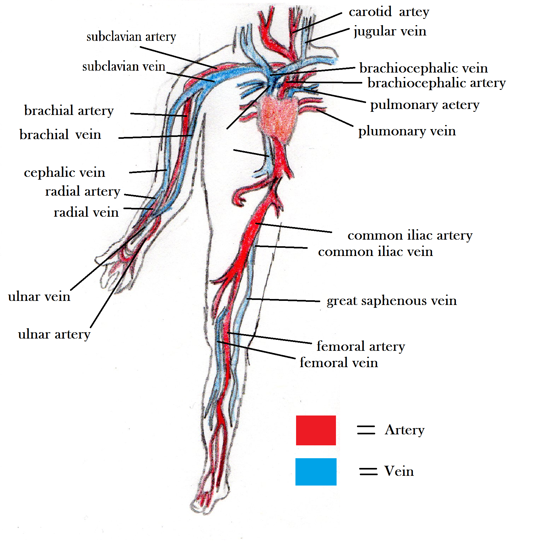
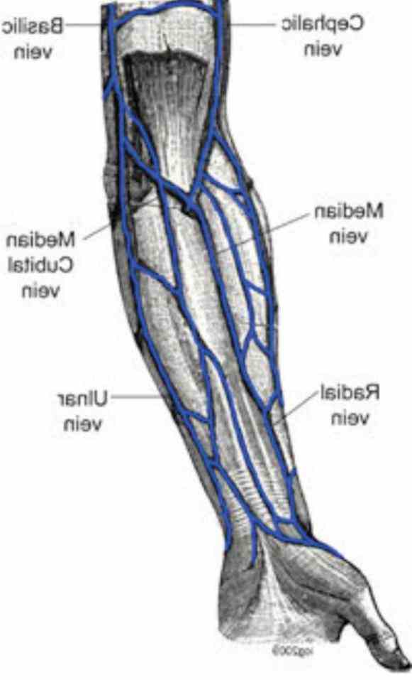
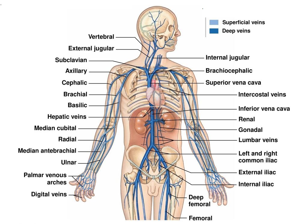
The venous system of the upper limb functions to drain deoxygenated blood from the hand, forearm and arm back towards the heart. Veins of the upper limb are divided into superficial and deep veins . The main superficial veins of the upper limb include the cephalic and basilic veins.

Pin by Emily Joy on Nurse life Nursing school survival, Nursing
Arteries and Veins of the Arm. Create healthcare diagrams like this example called Arteries and Veins of the Arm in minutes with SmartDraw. SmartDraw includes 1000s of professional healthcare and anatomy chart templates that you can modify and make your own. 13/71 EXAMPLES. EDIT THIS EXAMPLE.
Veins And Arteries Diagram exatin.info
Saphenous Vein Mapping Ultrasound. Your doctor has requested an ultrasound of veins in your legs. Ultrasound is a procedure that uses sound waves to "see" inside your body. This procedure is performed to create a "map" of your leg veins for the surgeon in preparation for various procedures that will include bypass graft surgery (replacing.

arm vein diagram Google Search Arm veins, Superficial veins, Human
This procedure is used to evaluate the veins in your arms for use during dialysis surgery or arterial bypass surgery in your arm or leg. At the Cedars-Sinai S. Mark Taper Foundation Imaging Center, we have a specialized team of physicians and technologists who are experts in ultrasound technology.
Anatomy Of The Veins In The Arm
The function of the basilic vein is to drain the blood from portions of your hand and arm so it can go back to the heart and lungs to be oxygenated and pumped out again. The dorsal venous network of the hand drains the blood from the palm of your hand and sends it upward to the basilic vein. Small branches of the basilic vein transport blood.

Veins of the Forearm TrialExhibits Inc.
The brachial vein (deep vein) accompanies the brachial artery in the region of the arm. It is formed by the unification of the ulnar and radial veins at the elbow. The basilic vein joins the brachial vein and becomes the axillary vein at the inferior border of the teres major muscle. At its terminal part the axillary vein is joined by the.

Download scientific diagram The superficial veins of the forearm and
Overview The arms are the upper limbs of the body. They're some of the most complex and frequently used body parts. Each arm consists of four main parts: upper arm forearm wrist hand Read on to.

Cephalic vein Wikipedia
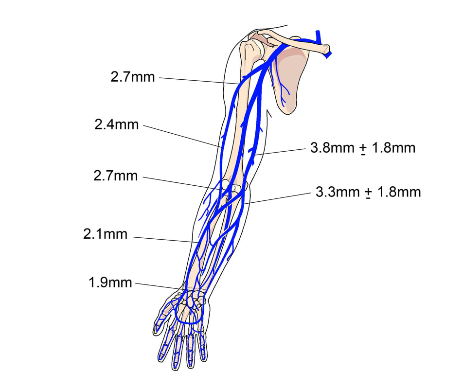
Upper Extremity Venous Mapping. Clinical Protocol. Place proximal blood pressure cuff or tourniquet (whenever possible) prior to making venous diameter measurements. Wait at least 2 minutes after blood pressure cuff placed before taking measurements. Device can be removed for small rest and then replaced during case if patient becomes.

Pin by Brenna on Get Schooled. Anatomy and physiology, Phlebotomy
In the upper arm, the basilic and cephalic veins are the major routes for superficial venous drainage, with ultimate runoff into the deep system (Figs. 77-4 and 77-5). The basilic vein is typically larger than the cephalic vein, coursing medial to the biceps brachii. The smaller cephalic vein courses lateral to the biceps brachii.

Vein Mapping. Hellllllo Nurse Pinterest
The brachial veins are located in the arm proper, the area between the shoulder and the elbow, and run alongside the brachial artery. The brachial veins work in reverse from the brachial artery. The ulnar and radial veins form a junction at the location where the brachial veins begin. The teres major muscle has an inferior border where the.

Pin by susan meyer on Vascular Access for Meds/Fluids/Nutrition Arm
What are the benefits vs. risks? What are the limitations of Venous Ultrasound Imaging? What is Venous Ultrasound Imaging? Ultrasound imaging is a noninvasive medical test that helps physicians diagnose and treat medical conditions. It is safe and painless. It produces pictures of the inside of the body using sound waves.
Veins Types, Venous System & Clinical Significance » How To Relief
Vein mapping is a technique performed with an ultrasound probe that uses sound waves (doppler) technology to view or "map" all of the veins under the skin on the arms or legs. It allows the doctor to see the size, depth, and flow of blood in these veins and allows for better treatment planning.

Anatomy Of The Veins In The Arm
Fifty-two adult healthy volunteers were evaluated for superficial vein diameter, brachial artery flow and diameter in the lower third of non-dominant arm by a dedicated vascular access radiologist blinded for the identification of the participants. Each participant was scheduled for three evaluations one week apart.

veins of the upper arm Veins, Upper arms, Iv therapy
Anatomy Basic deep venous anatomy of the arm. Basic superficial venous anatomy of the arm. Deep Veins of the Neck & Shoulder The red line shows the subclavian vein origin scan plane. Ultrasound Doppler of the Subclavian vein origin. Ultrasound of the Jugular vein pre & post compression.

Cephalic vein Wikipedia (With images) Greys anatomy book, Arm veins
Some of the veins in the arm include: Dorsal venous network: This web of veins extends across the back of the hand. Superficial veins: As their name implies, these veins are close to the.
forearm veins anatomy
Arterial and venous mapping, commonly called vein mapping, is an ultrasound test that takes pictures of your arteries and veins (blood vessels). This test creates a "map" of your blood vessels that can be used to help guide some medical procedures or to check for certain health conditions. Advertisement